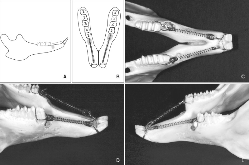
Figure 1.
Study design and device. A, Buccal view; B, occlusal view of a schematic drawing. Right, drilled socket; left, extraction socket. The drilled socket was created mesial to the first premolar on the right side while the left first premolar was extracted. Clinical photos of rabbit mandible showing the occlusal view and the buccal views of the intentional and extraction sockets (C, D, and E, respectively).

