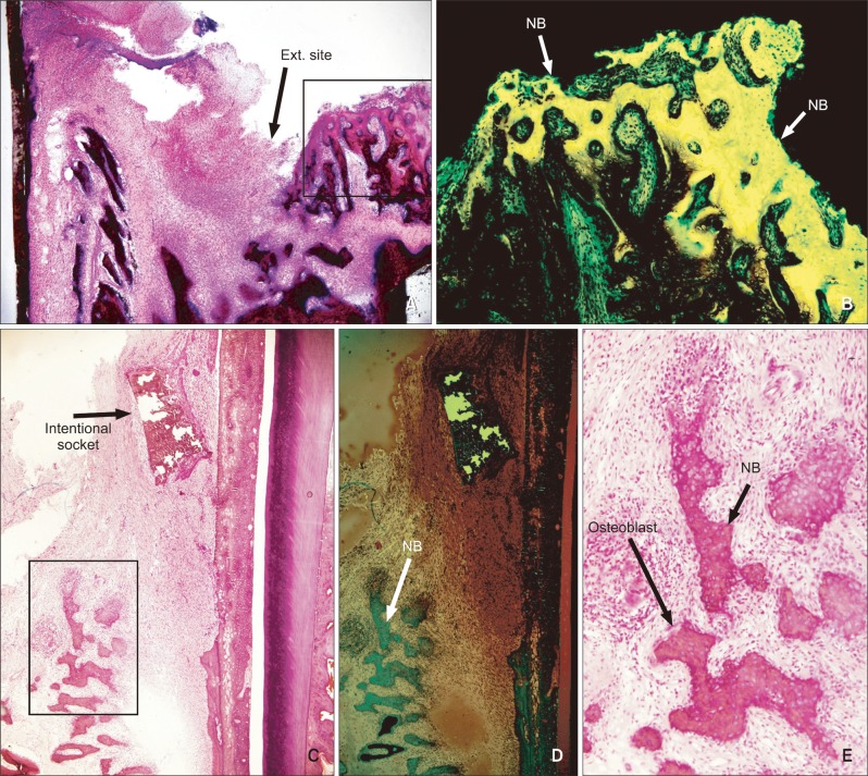
Figure 3.
Representative histological features of the intentional socket site in an undecalcified section. The extraction (Ext.) socket group (A, ×40) shows newly formed bone (NB) and host bone after 2 weeks. B, Fluorescent microscope photo of A. At week 2, the intentional socket group (C, ×40) shows new bone under fluorescence microscopy (D, ×100) and active osteoblasts (E, ×100).

