Figure 5.
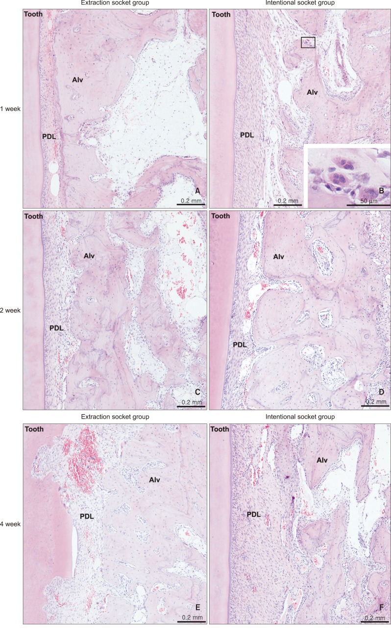
Microphotograph of the periodontium with H&E staining. A, C, E, Extraction socket at weeks 1, 2, and 4, respectively; B, D, F, intentional socket at weeks 1, 2, and 4. PDL, Periodontal ligament; Alv, alveolar bone. Note the enlarged PDL space in B, D, and F, and the increased number of osteoclasts on some resorbed bone surfaces, especially at week 1 in the intentional socket (B), which might have caused the enlarged PDL space by its increased activity. Root resorption can be noticed in E. The figure suggests elevated alveolar bone resorption in both groups from week 1 - 4, particularly in the intentional socket group.

