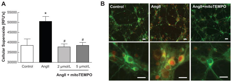
Figure 2. Cellular and mitochondrial superoxide scavenging by mitoTEMPO in Ang II-treated cultured neuron.
A, Cellular superoxide stained by dehydroethidium. Relative fluorescence were detected by microplate reader. *P<0.05 vs control, #P<0.05 vs Ang II. B, Representative images of neurons. mitoSOX (red) staining is used to detect mitochondrial superoxide and mitoTracker (green) is used for mitochondrial localization. Scale bars=50μm.

