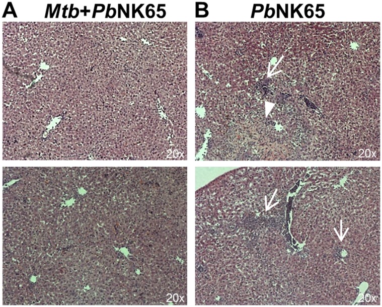
Figure 3. PbNK65 associated liver damage is reduced in co-infected mice.
C57BL/6 mice were aerosol infected with M. tuberculosis H37Rv (100 CFU/lung) and 40 days later challenged with PbNK65 sporozoites by mosquito bite. Control mice were infected with M. tuberculosis or PbNK65 alone, respectively. Representative H&E stains of liver sections 13 days after co-infection. Note, that PbNK65 infection caused periportal inflammation (B; arrows) and tissue necrosis (B; arrowhead) which was reduced in livers of co-infected animals (A).

