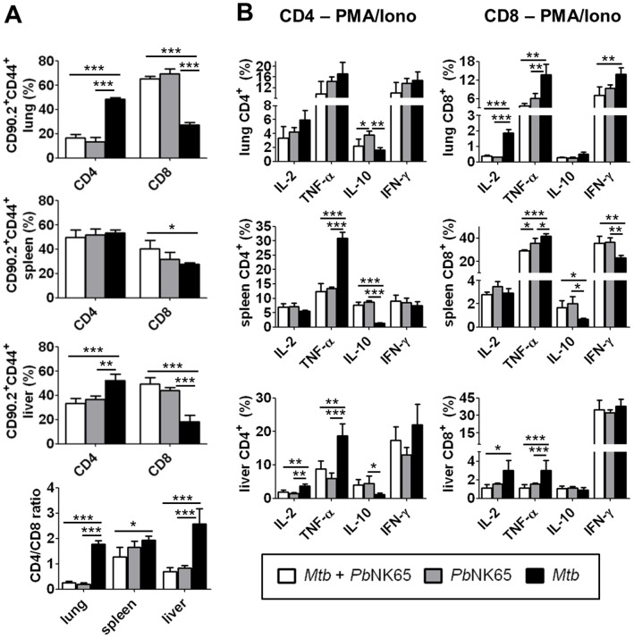
Figure 4. PbNK65 co-infection alters T cell responses in M. tuberculosis infected mice.
C57BL/6 mice were infected by aerosol with M. tuberculosis H37Rv (100 CFU/lung) and challenged with PbNK65 sporozoites by mosquito bite 40 days later. Control mice were infected with M. tuberculosis or PbNK65 alone, respectively. A) 12 days upon PbNK65 infection, lungs, spleens and livers were analyzed for the presence of CD44 positive CD4 and CD8 effector T cells by flow cytometry. B) Whole lung and spleen lysates and purified liver lymphocytes were re-stimulated in vitro with PMA/Iono (50 ng/ml, respectively) and analyzed by flow cytometry for the presence of IL-2, TNF-α, IL-10 or IFN-γ producing CD4 and CD8 T cells. Results are shown as means ± SD (n = 3–5). Data from one out of two independent experiments are shown. Statistical analysis was performed by ANOVA (*p<0.05; **p<0.01; *** p<0.001).

