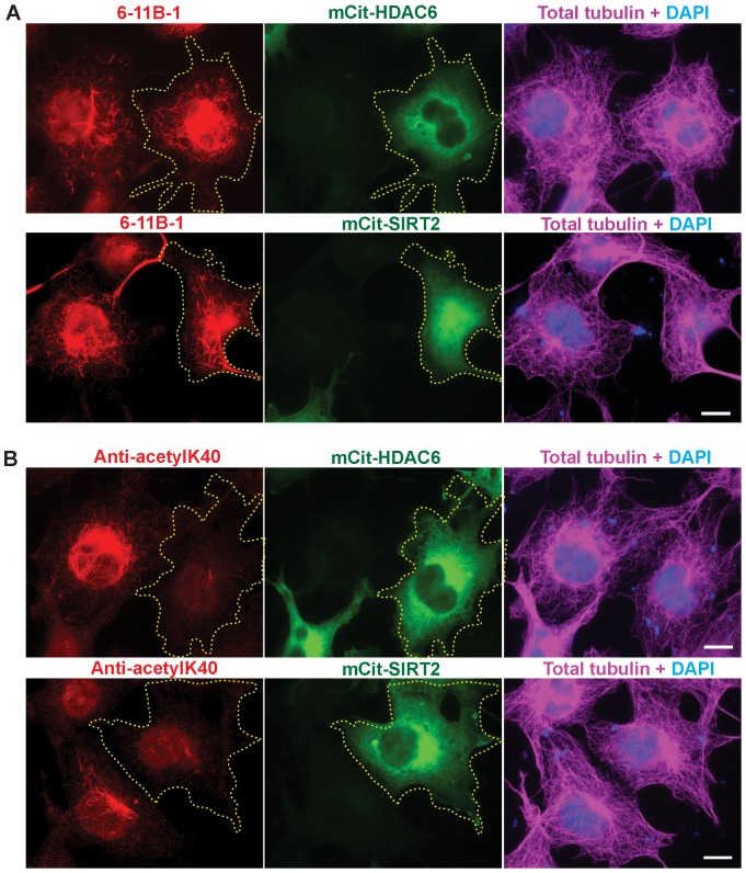
Figure 5. Monoclonal (6-11B-1) and polyclonal (anti-acetyl-K40) antibodies differ in their ability to recognize deacetylated microtubules in cells.
COS7 cells expressing the deacetylases mCit-HDAC6 or mCit-SIRT2 (green) were fixed and double stained using A) monoclonal 6-11B-1 (red) and total tubulin (magenta) antibodies or B) polyclonal anti-acetyl-K40 (red) and total tubulin (magenta) antibodies. Transfected cells are indicated by the yellow dotted outline. Scale bars, 20 µm.

