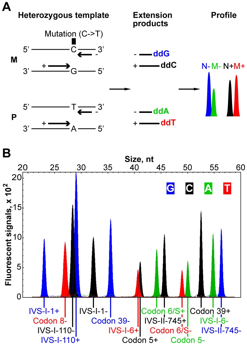
Figure 1. Developing the single-nucleotide primer extension assay.
(A) Principle of the single-nucleotide primer extension method illustrated through analysis of a sample carrying a point mutation of interest. Four template DNA strands from the maternal (M) and paternal (P) chromosomes are shown (variable nucleotide lettered). The template is interrogated by two extension primers (thick arrows) giving rise to normal and mutant extension products and peaks. ‘+’ and ‘−’ indicate strand specificity of the primers; the fluorescently labeled nucleotides incorporated into extension products are bold and colored as they appear on the electropherogram. N+, normal peak generated from ‘+’ primer; M+, mutant peak generated from ‘+’ primer; N−, normal peak generated from ‘−’ primer; M−, mutant peak generated from ‘−’ primer. (B) Normal DNA electropherogram profile obtained with the optimized primer set: primer extension product peaks are labeled with the corresponding primer names as in Table 2.

