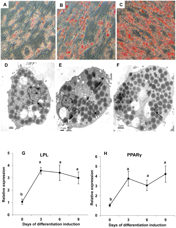
Figure 2. Micrographs of large yellow croaker adipocytes differentiated in culture.
The cells were induced to differentiate into adipocytes and stained with oil red O at (A) day 3, (B) day 6 and (C) day 9 (×200) after induction. Electron micrographs of yellow croaker preadipocytes differentiated in culture at (D) day 3, (E) day 6 and (F) day 9 after induction. Bars: D, E = 1 µm, F = 2 µm. Arrows points to lipid droplets.

