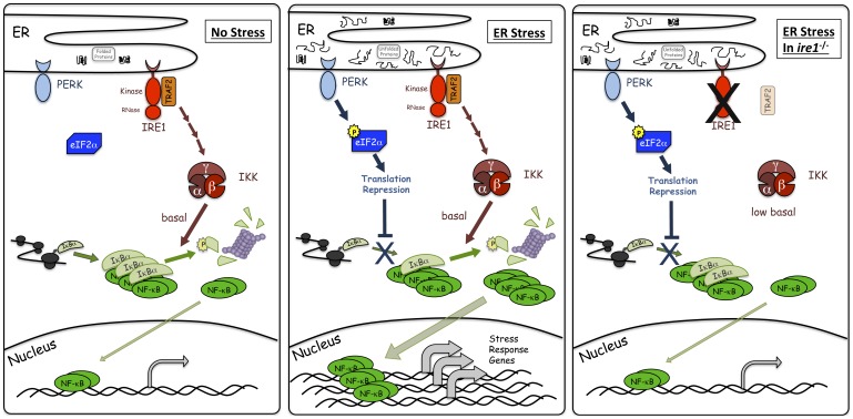
Figure 6. Model for NF-κB activation during ER stress.
Under unstressed conditions, IκBα is being synthesized, binds, and inhibits NF-κB. IRE1, through TRAF2, is maintaining basal IKK activity which is responsible for phosphorylating a subset of IκBα leading to proteosomal degradation and basal NF-κB activity. However, most of the NF-κB is sequestered by IκBα. During ER stress, PERK phosphorylation of eIF2α leads to translation repression which then prevents synthesis of new IκBα and contributes to a decrease in IκBα levels and corresponding increase in free NF-κB levels. Additionally, basal IKK activity is responsible for phosphorylating and degrading the existing IκBα, including IκBα bound to NF-κB, causing a more dramatic decrease in IκBα levels resulting in an even greater amount of free NF-κB. Free NF-κB can then translocate to the nucleus to assist in transcriptional activation of stress response genes. During ER stress in cells with decreased basal IKK, such as ire1 −/− cells, basal IKK is considerably reduced. PERK mediated translation inhibition alone is unable to reduce IκBα levels enough to allow for a significant amount of free NF-κB. Thus, combined inputs from both PERK are IRE1 are required for full activation of NF-κB during ER stress. It should be noted that the possibility remains that additional element(s) beyond both PERK induced translation repression and IRE1 regulation of basal IKK/IκBα stability, may also contribute to overall activation of NF-κB during ER stress.

