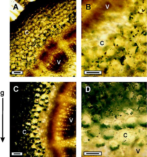
Figure 5.
Light micrographs of chloroplast distribution in cortical cells of the stem-bending zone as seen in cross-sections prepared from vertical (A and B) or gravistimulated (90°; C and D) cv Potomac Royal spikes. Cross-sections of the stem-bending zone taken from vertical or gravistimulation spikes were stained with KI/I2 solution and examined under the microscope. c, Cortex; v, vascular cylinder. Bars = 25 μm.

