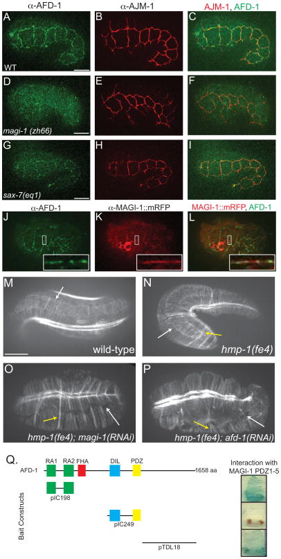
Figure 4. MAGI-1 influences localization of AFD-1.
(A–C) Immunostaining of AFD-1 in wild-type embryos shows AFD-1 accumulates at cell junctions (A), but there is little overlap with AJM-1 (C). (D–F) AFD-1 does not accumulate at cell junctions in magi-1 null embryos (D), even though AJM-1 still localizes to junctions (E). (G–I) Consistent with the interaction between SAX-7 and MAGI-1, AFD-1 accumulation at junctions in sax-7(eq1) embryos is reduced (G). (J–L) Immunostaining against MAGI-1::mRFP (K) and AFD-1 (J) reveals a high degree of colocalization between the two proteins (L, inset). Scale bar is 10 μm. (M–P) Phalloidin staining of wild-type embryos shows accumulation of actin and ordered CFBs anchored at the junction (white arrow, M). (N) CFBs in hmp-1(fe4) embryos are also anchored at the junction (white arrow); however, some of the CFBs are clumped together (yellow arrow). Similar staining in hmp-1(fe4); magi-1(RNAi)(O) and hmp-1(fe4); afd-1(RNAi) (P) embryos reveals a loss of junctional proximal actin and CFBs are completely detached from the junction (white arrows). There are also several clumps of CFBs (yellow arrows). (Q) In a directed yeast two-hybrid test, the PDZ domains in MAGI-1 interacted with the RA domains and the C-terminus, but not the DIL-PDZ domains of AFD-1. Scale bars are 10 μm.

