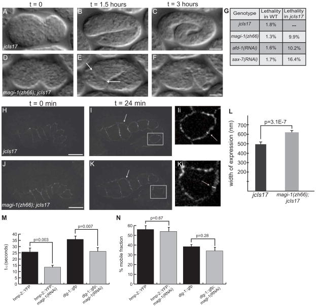
Figure 5. MAGI-1 helps to partition junctions during development.
(A–C)jcIs17 embryos enclose (A) and elongate as wild-type embryos do, reaching approximately 4-fold before they hatch (C). (D–F) magi-1(zh66); jcIs17 embryos enclose ventrally, but the anterior epidermis does not complete enclosure. When elongation starts, anterior cells spill out of the opening in the epidermis (arrows, E). Scale bars are 10 μm. (G) Depleting magi-1, afd-1 or sax-7 in jcIs17 embryos causes similar non-additive increases in lethality, also primarily due to anterior enclosure defects. (H–K) At the onset of elongation, jcIs17 (H) and magi-1(zh66); jcIs17 (J) embryos look indistinguishable with respect to HMP-1::GFP. As elongation progresses, HMP-1::GFP expression is expanded at seam-dorsal and seam-ventral boundaries in magi-1(zh66); jcIs17 embryos (arrows, Ii, Ki; white lines indicate width of junctional material). Scale bars are 10 μm. (L) magi-1(zh66); jcIs17 embryos exhibit an expanded area of HMP-1::GFP expression (618±29 nm, mean ± SEM, n=10) in seam cells compared to wild-type embryos (493±49 nm, n=13). Significance was calculated using a two-tailed Student’s t-test. (M) magi-1(RNAi) decreases the half-life (t1/2) of fluorescence recovery after photobleaching in embryos expressing hmp-2::yfp or dlg-1::gfp. (N) magi-1(RNAi) does not significantly affect the mobile fraction of either HMP-2::YFP or DLG-1::GFP.

