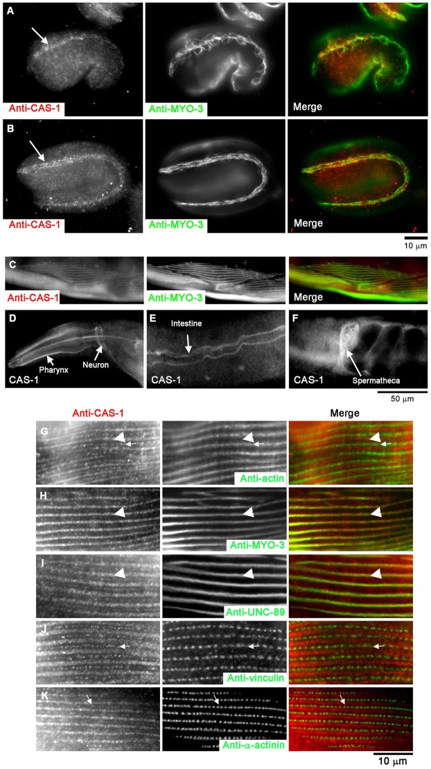
Fig. 1.
Expression and localization patterns of CAS-1 in C. elegans embryos and adult worms. (A,B) Expression patterns of CAS-1 in embryos at the 1.5-fold (∼420-min old; A) and 2-fold (∼450-min old; B) stages. Embryos were fixed and stained with anti-CAS-1 (left panels) and anti-MYO-3 (a marker for the body wall muscle; middle panels). Merged images are shown in the right panels (CAS-1 in red and MYO-3 in green). CAS-1 was expressed predominantly in the body wall muscle as indicated by arrows in A and B. (C–F) Expression patterns of CAS-1 in adult worms. (C) Adult worms were fixed and stained with anti-CAS-1 (left panel) and anti-MYO-3 (middle panel), and a region of the body wall muscle is shown. A merged image is shown in the right panel (CAS-1 in red and MYO-3 in green). Immunostaining of CAS-1 was also detected in the pharynx (D), the neurons (D), the intestinal lumen (E), and the spermatheca of the somatic gonad (F). (G–K) Sarcomeric localization of CAS-1 in C. elegans adult body wall muscle. Adult worms were fixed and stained with anti-CAS-1 (left panels) and anti-actin (G, middle), anti-MYO-3 (H, middle), anti-UNC-89 (I, middle), anti-vinculin (J, middle) or anti-α-actinin (K, middle) antibodies. Merged images are shown in the right panels (CAS-1 in red, and actin, MYO-3, UNC-89, vinculin and α-actinin in green). Arrowheads in G–I indicate positions of the M-lines, and arrows in G, J and K indicate positions of CAS-1 spots in the I-bands.

