Fig. 7.
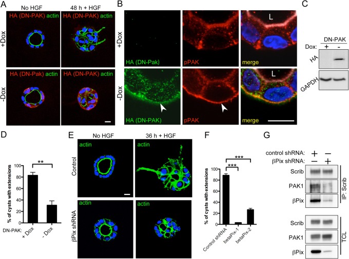
PAK and βPix are required for HGF-mediated cyst extension formation. (A) Tet-off-regulated expression of an HA-tagged DN-PAK (K299R) kinase-dead construct. Cysts were cultured for 5 days in Matrigel and in the presence or absence of doxycycline (+/– Dox) to regulate expression. Cysts were treated with 50 ng/ml HGF for 48 hours to induce extensions. Anti-HA immunofluorescence staining (red) of fixed cysts was performed to monitor DN-PAK expression, and fluorescently labeled phalloidin (green) was used to visualize actin. Nuclei were stained with Hoechst (blue). (B) Magnification of DN-PAK-expressing cysts showing basal localization of DN-PAK. pPAK colocalizes at the basal surface with DN-PAK, but not in control cysts (arrows). Merged images also show actin, in white, marking the apical surface. L, lumen. (C) Dox-regulated expression of DN-PAK was also monitored by western blot analysis and anti-GAPDH was used as a cell lysate loading control. (D) The percentage of cysts with one or more extensions in the presence or absence of Dox was quantified. (E,F) 4-day-old cysts of control cells or cells infected with βPix lentiviral shRNA were induced with HGF (E) and the ability of cysts to form extensions was quantified (F). Dunnett's multiple comparison test was used. (G) IPs of Scrib in control- or βPix-shRNA-treated cells. Less PAK is associated with Scrib following βPix RNAi. **P<0.01, ***P<0.001. Scale bars: 10 µm.

