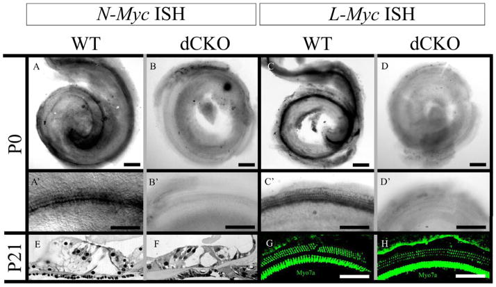
Figure 1. The complete loss of N-Myc and L-Myc in inner ear hair cells does not affect overall organ of Corti organization or hair cell cytoarchitecture.
N-Myc (A) and L-Myc (C) are expressed in the cochlea after birth. Their expression is prominent in cochlear hair cells (A′ and C′). Using Atoh1-Cre, N-Myc (B) and L-Myc (D) were completely removed from inner ear hair cells, specifically noted is their absence in the organ of Corti (B′ and D′). However, this absence had no noticeable effect on overall architecture of the organ of Corti as three rows of outer hair cells and one row of inner hair cells were present throughout the cochlea along with the proper arrangement of supporting cells seen in epoxy resin sections of WT (E) and dCKO (F) mice as well as with myosin VIIa immunohistochemistry of WT (G) and dCKOs (H), shown near the middle turn. Scale bar = 100μm.

