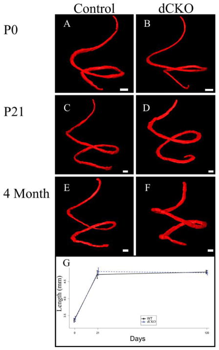
Figure 2. The loss of N-Myc and L-Myc does not change the normal morphologic development of the organ of Corti.
Myosin VIIa positive hair cells and rhodamine stained organ of Corti nuclei were segmented at P0 in WT (A) and dCKO mice (B), P21 (C and D) mice, and four months of age (E and F) mice. Segmented hair cells were three dimensionally reconstructed to provide an accurate measurement of the length of the organ of Corti at these three time points. From P0 to four months of age, the organ of Corti lengthened (G); however, this increase in length was similar between WT and dCKO mice. Scale bar = 100μm.

