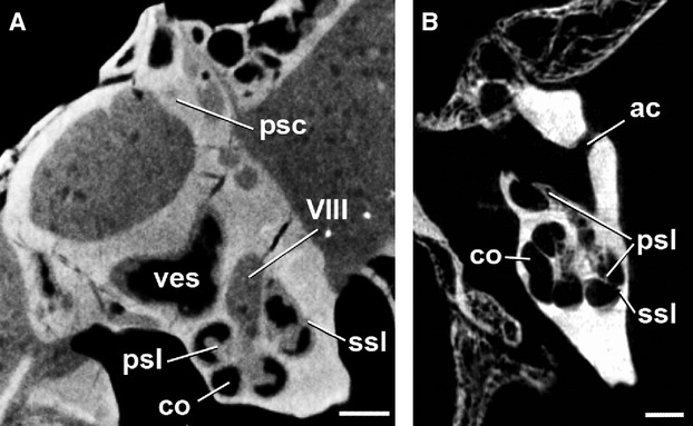
Fig. 4.

Transverse CT image through the ear region of the skull of (A) Diacodexis ilicis (AMNH VP 16141), (B) Moschiola meminna (UM2 58V), showing the internal anatomy of the left petrosal. The secondary osseous spiral lamina is visible as a faint structure projecting from the meatal wall of the cochlear canal. Scale bar: 5 mm. ac, cochlear aqueduct; co, cochlear canal; psc, posterior semicircular canal; psl, primary osseous spiral lamina; ssl, secondary osseous spiral lamina; ves, vestibule; VIII, opening for vestibulocochlear nerve (Cranial Nerve VIII).
