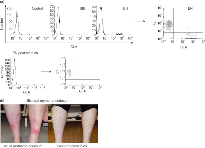
Fig. 6.

Aberrant expression of cutaneous lymphocyte-associated antigen (CLA) on γδ T cells in erythema nodosum. (a) Fluorescence activated cell sorter (FACS) histograms demonstrating proportions of circulating γδ T cells in healthy controls (0·8 ± 0·3%, n = 9), active inflammatory bowel disease (IBD) (1·2 ± 0·4%, n = 15) and erythema nodosum (EN) with inactive IBD (9·6% and 5·7%, n = 2). Bottom row: EN post-steroids (1·3%, n = 1; pre-steroids was 9·6%) expressing CLA. Histograms are representative of several independent experiments performed with similar results, and were compared to isotype-matched controls; FACS dot-plot demonstrating proportions of circulating γδ T cells in EN co-expressing gut-homing marker β7 and skin-homing marker CLA, expressing β7 only, or expressing CLA only. All plots were compared to isotype-matched controls. (b) Photographs of shins of EN patient pre- and post-corticosteroids.
