
Fig. 4.
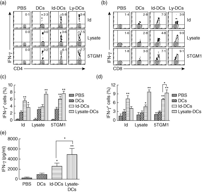
Cellular immune responses in mice receiving dendritic cell (DC) vaccines. Flow cytometry analyses showing the expression of interferon (IFN)-γ in gated CD4+ (a) or CD8+ (b) T cells in response to idiotype (Id) protein, tumour lysate (lysate) or irradiated 5TGM1 myeloma cells (5TGM1). Values in each dot-plot represent the percentages of CD4+ or CD8+ T cells expressing IFN-γ. Summarized data for percentages of IFN-γ-expressing CD4+ (c) or CD8+ (d) T cells from three different experiments are shown. In these studies, tumour-free mice (three per group) were vaccinated with three weekly subcutaneous injections of Id-keyhole limpet haemocyanin (KLH)-pulsed DC vaccine (Id-DCs) or tumour lysate-KLH-pulsed DC vaccine (lysate-DCs). Phosphate-buffered saline (PBS) and unpulsed DCs (DCs) served as controls. One week later, splenocytes were isolated, pooled and restimulated with Id protein, tumour lysate or irradiated 5TGM1 myeloma cells for 24 h. (e) Amount of secreted IFN-γ by tumour-specific T cells in splenocytes of mice that were restimulated with irradiated 5TGM1 myeloma cells for 3 days. Cytokine in cell culture media was quantified by enzyme-linked immunosorbent assay (ELISA). Representative results from one of three independent experiments are shown. The error bars represent stand deviations of three independent experiments. **P < 0·01, compared with PBS or unpulsed DC controls.