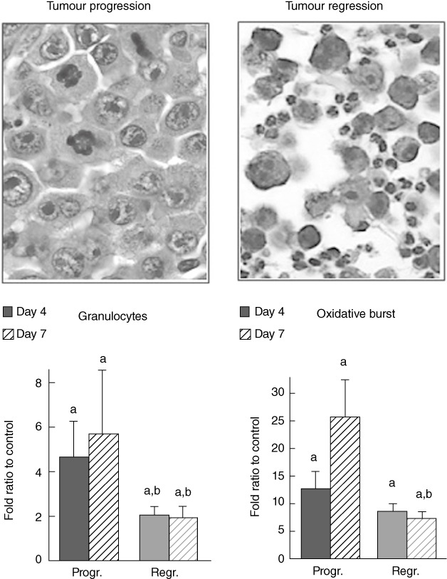
Fig. 2.

The involvement of granulocytes in tumour growth. Distribution of peripheral blood granulocytes [mean ± standard error (s.e.) per group], granulocyte oxidative burst (1O2 production measured by chemiluminescence assay) during tumour progression or regression (mean ± s.e. per group) and representative histological image of tumour transplantation site taken on day 4 in tumour progression or regression tissue samples (both ×400, haematoxylin and eosin) are shown. (a) Significance P < 0·05 in comparison to healthy controls, (b) significance P < 0·05 in comparison to samples obtained from tumour progressing animals.
