
Fig. 1.
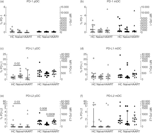
Expression of programmed death (PD) receptors by plasmacytoid dendritic cells (pDC) and myeloid DC (mDC) from HIV-1+ patients. Peripheral blood mononuclear cells (PBMC) from healthy controls (circles), therapy-naive (squares) and therapy-treated patients (triangles) were collected, stained and analysed by flow cytometry. Expression of PD-1 (top plots), PD-L1 (middle plots) and PD-L2 (bottom plots) by pDC (left plots) and mDC (right plots) are shown. Light symbols indicate the percentages of cells expressing the specific markers (left y-axes), whereas dark symbols represent the mean fluorescence intensity of each receptor (right y-axes). The lines in the middle show the median value, whereas error bars represent the 25th and 75th percentiles. Statistically significant P-values are shown (Mann–Whitney U-test).