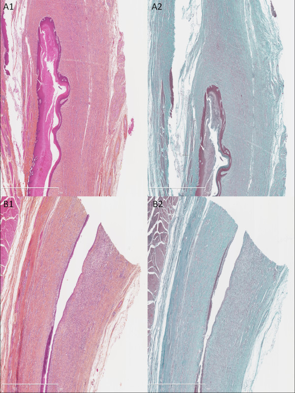
Figure 4.
Histological analysis of the resected esophagus with predominant fibrotic pattern. Presence of a neoepithelium, as a single layer of immature epithelial cells, a thin granulation tissue with moderate cell density, and important thickness of cicatricial fibrosis infiltrating the remaining submucosal and the muscular layer. (A1) Untreated swine, hematoxylin-eosin-safran (HES), original magnified x25. (A2) Untreated swine, Masson’s trichrome, original magnified x25. (B1) Treated by N-acetylcysteine (NAC), HES, original magnified x25. (B2) Treated by NAC, Masson’s trichrome, original magnified x25.

