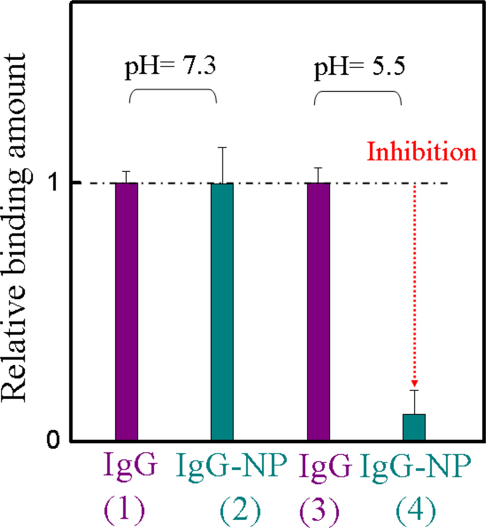
Figure 7.
Interaction between the Fc fragment of IgG and protein A at pH 7.3 and 5.5 in 35 mM phosphate buffer, before and after addition of NP7. The experimental design is similar to Figure 6a. Whole IgG was used to functionalize the QCM cell. The color of the column in the graph corresponds to the color of the arrowhead in Figure 6a. Cyan column: the frequency change was caused by protein A binding to the Fc domain of IgG in the presence of NP7 at pH 7.3 (2) and 5.5 (4), respectively. Purple column: control experiment under the same conditions except omitting the introduction of NPs at pH 7.3 (1) and 5.5 (3). Relative binding is defined as comparing the binding amount of the NP to that of protein A for whole IgG. Standard deviations were calculated from three independent measurements.

