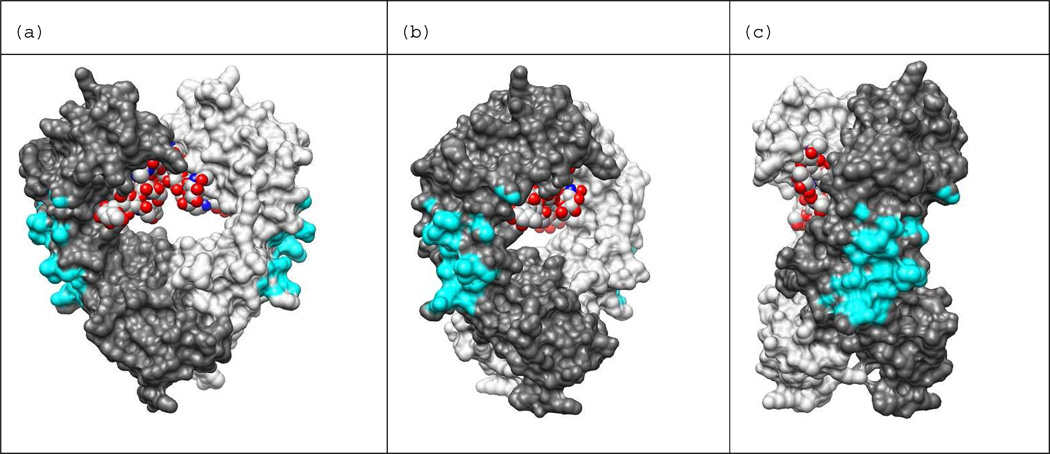
Figure 8.
Protein A binding site shown on IgG1-Fc dimer. Binding site atoms are colored cyan. An Fc atom is considered in contact with protein A if it is within 5 Å of any heavy atom of protein A. The holo structure PDB ID: 1FC2 was used to calculate distances between Fc and protein A. The N-terminus of Fc is at the top of the view and C-terminus is at the bottom. Chain H is colored dark gray, chain K is colored light gray, and the carbohydrates bound in the Fc dimer core (red) are shown in the space filled representation. Images generated from PDB ID: 1HZH with UCSF Chimera.42 The view in (b) is rotated 45 degrees about the vertical axis with respect to the view in (a), and the view in (c) is rotate 90 degrees with respect to (a).

