Figure 10.
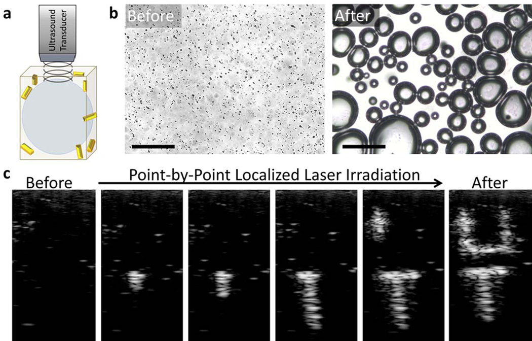
Ultrasound contrast enhancement in vitro. (a) Depiction of the gas phase of a PAnD after laser triggered vaporization has occurred. These microbubbles provide significant acoustic impedance mismatch between the PFC gas and the surrounding environment. (b) Optical images of a hydrogel with PAnDs before laser exposure and after laser exposure. Individual droplets are expected to create bubbles approximately 5 time the diameter of the original droplet. The larger bubbles are due to rapid coalescence of smaller bubbles. Scale bars represent 50 µm. (c) Sequential US frames captured as the laser irradiation produced desired pattern in the phantom. The image before laser irradiation illustrates that the ultrasound field alone does not activate PAnDs (i.e., does not initiate the liquid-to-gas transfer of the PFC). As PAnDs are irradiated with laser beam at corresponding positions, the microbubbles are locally triggered, resulting in ultrasound contrast enhancement. Each individual spot is approximately 1 mm, with the final letters standing 1.2 cm tall and 0.5 cm wide. Images are in 20 dB scale. Adapted from ref.176 with permission.

