Figure 1.
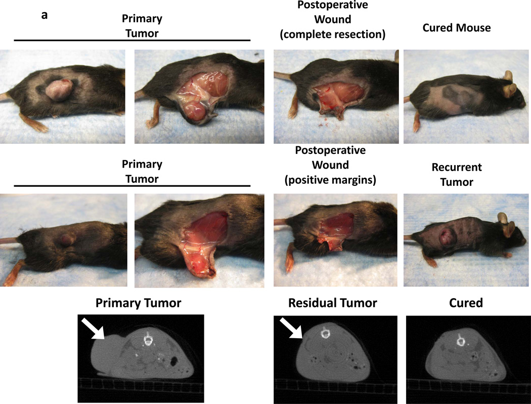
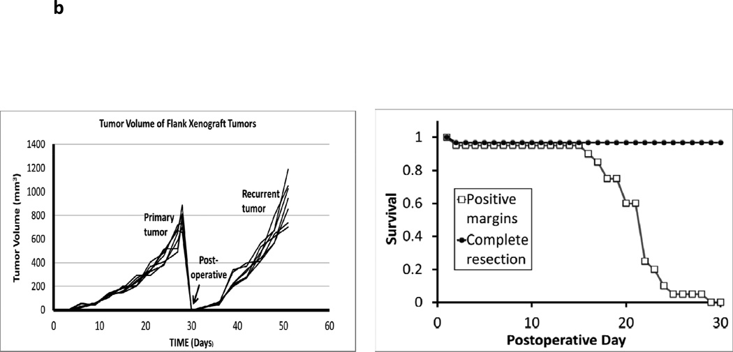
A surgical model for local recurrence of disease following surgery. Syngeneic immunocompetent mice were injected with 1.5e6 TC1 tumor cells into the right flank (not over the liver) and developed large tumors (>800 mm3). When the tumors measured 800mm3, they were either partially resected with positive margins or completely removed. In the partial resection group, the surgeon used a #15 scalpel to sharply divide the tumor and leave the smallest possible residual nodule (<5%) that was technically feasible (ranged 2 to 4 mm in diameter). This tumor deposit was typically left attached to either the skin or underlying muscle. (A) A representative animal from each group is shown. Two independent observers first visually inspected the wound, and then palpated the wound in order to determine which animals had residual disease versus curative surgery. Animals (n=20) without obvious tumor to independent observers underwent microCT scans to determine if residual disease could be radiographically located. White arrows designate flank tumors. (B) Following surgical intervention, all animals with (i) positive margins and (ii) total resections were monitored for recurrence of their flank tumors. Tumor volume vs time and Kaplan Meier survival curves for a typical flank TC1 (n=20) experiment are depicted.


