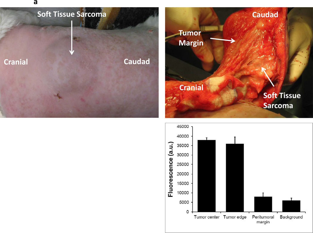
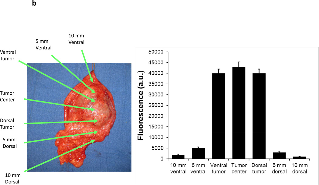
Figure 5.
A canine with a spontaneous abdominal wall sarcoma underwent image guided surgery. After ICG injection, (A) a canine abdominal was opened to reveal a well demarcated soft tissue sarcoma. The primary tumor was imaged in 8 radial directions from the center of the tumor and fluorescence averaged. The tumor bed was imaged prior to wound closure. (B) After resection, ex vivo fluorescence measurements were again performed.


