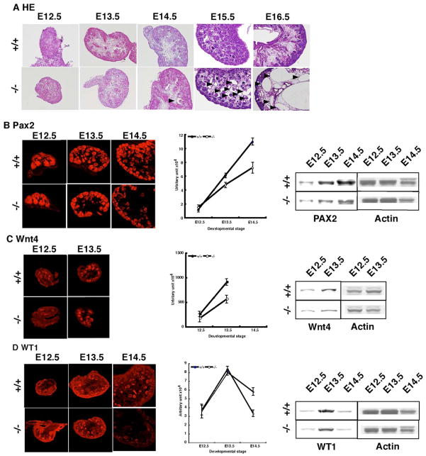
Figure 1. Precise characterization of nephrogenesis in Pkd−/− mice.
To examine whether Pkd1−/− mice display an impairment of mesenchymal differentiation, we performed immunohistochemistry using PAX2, WNT4 and WT1 as markers. (A) H-E stain revealed the formation of cysts to be remarkable at E15.5 as previous reports (arrowhead)[16, 20]. A small number of cysts were was detectable from E14.5 (arrowhead). There were no obvious cyst before E13.5. (B) We first analyzed PAX2 expression. At E12.5, the expression was completely normal, but after E13.5 it was significantly reduced compared that in wild type littermates. To quantitate the level precisely, we measured PAX2 expression in serial sections as shown in Supplemental Figure1 and Supplemental Table 1. A Western bloting is shown on the right side. (C) We also examined WNT4 expression. Similarly, the expression was reduced from E13.5. The expression of WNT4 was markedly downregulated thereafter, and was not detectable after E14.5. A Western bloting is shown on the right side. (D) Finally, we examined the expression of WT1. The expression was relatively normal by E13.5, but was more rapidly downregulated in Pkd1−/− mice at E14.5.

