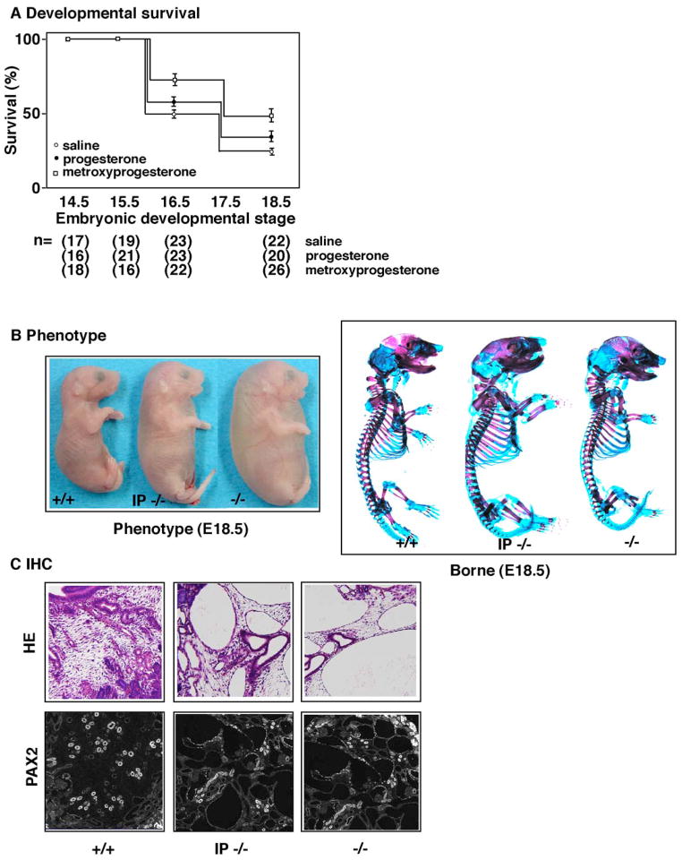
Figure 3. Improvement of phenotype in Pkd−/− mice on the intraperitonial injection of progesterone and a derivative.
To examine whether progesterone and its derivatives are effective against the phenotype of Pkd1−/− embryos, we injected them into the intraperitoneal space of pregnant females of Pkd1+/− mice. (A) The rate of survival was examined at a given point in gestation. Most of the Pkd1−/− embryos died before E18.5, whereas in the injected group, over 35% of Pkd1−/− embryos survived. The effect on the rate of survival rate was significant in medroxyprogesterone acetate, but less significant in progesterone. The total number of embryos examined is summarized at the bottom. (B) Surface examination revealed that Pkd1−/− embryos at E17.5 are edematous and pale compared to wild type controls (+/+), whereas Pkd1−/− embryos from injected groups (IP−/−) are improved compared to the uninjected group (−/−) (left panel). Quantitation of fluid volume is summarized at the bottom. Skeletal bone stain revealed an impairment of osteogenesis in Pkd1−/− embryos, whereas Pkd1−/− embryos from injected groups (IP−/−) are partially restored compared to the uninjected group (−/−) (right panel). Wild type (+/+) is shown at the left. (C) We examined H-E staining of Pkd1−/− embryos from injected groups (IP−/−) compared to the uninjected group (−/−), and found a partial effect on the formation of cysts. In these cases, expression of PAX2 was also improved. Wild type (+/+) is shown at the left.

