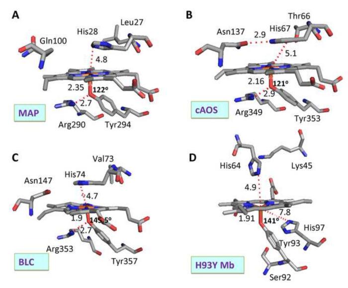
Fig. 1.
Active site structures of native ferric (A) MAP, (B) cAOS, (C) BLC and (D) ferric H93Y Mb mutant. In this comparison, the structures of the heme, the proximal tyrosinate ligand and several amino acid groups that are located near the heme at both the proximal and distal sides are illustrated and selected inter-atomic distances and Fe–O–C-Tyr bond angles are shown. These structures were drawn using PDB numbers 3E4W (MAP) [8], 1U5U (cAOS) [4], 8CAT (BLC) [9] and 1HRM (H93Y Mb) [10].

