
Figure 4.
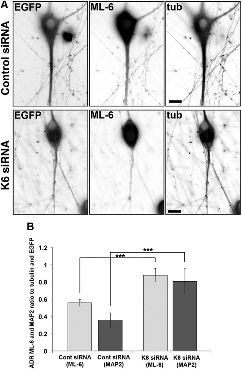
Depletion of kinesin-6 causes MAP2 and residual ML-6 immunolabeling to become redistributed in the neuron. SCG neurons were transfected with siRNA at plating and then with EGFP and control or kinesin-6 siRNA at 5 DIV using the CX1 device. Neurons were fixed in methanol at 7 DIV and double labeled with ML-6 and β-III tubulin (tub). Fluorescence images are presented in an inverted format that converts blacks to whites and whites to blacks and inverts gray levels proportionally. A, At 7 DIV, neurons transfected with control siRNA exhibited thick dendrite morphology, whereas neurons transfected with kinesin-6 siRNA only exhibited thin experimental dendrites. B, Quantification of the mean ADR in neurons double labeled with ML-6 and β-III-tubulin or ML-6 and MAP2 at 7 DIV. Data are represented as mean ADR (see Materials and Methods), in which ADR for ML-6 is significantly lower in control neurons than kinesin-6-depleted neurons. In control neurons, ML-6.ADR = 0.56 ± 0.04. In cultures depleted of kinesin-6, ML-6.ADR = 0.88 ± 0.08, (n = 30; p < 0.001). In control neurons, MAP2.ADR = 0.36 ± 0.08. In cultures depleted of kinesin-6, MAP2.ADR = 0.81 ± 0.15 (n = 20; p < 0.001).