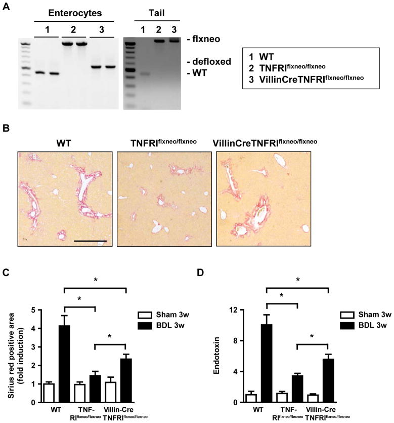
Figure 6. Reactivation of TNFRI on intestinal epithelial cells contributes to liver fibrosis.
(A) Reactivation of TNFRI on isolated colonic epithelial in VillinCreTNFRIflxneo/flxneo was analyzed by PCR (left panel). Tails from respective mice served as controls (right panel). (B–D) Wild type, TNFRIflxneo/flxneo and VillinCreTNFRIflxneo/flxneo mice underwent sham operation (n=3–4) or bile duct ligation (BDL; n=6 for C57BL/6, n=9–10 for TNFRIflxneo/flxneo and for VillinCreTNFRIflxneo/flxneo mice). (B and C) Collagen deposition was evaluated by Sirius red staining and quantitated by image analysis. Scale bar = 50μm. (D) Plasma endotoxin levels were measured. *p < 0.05.

