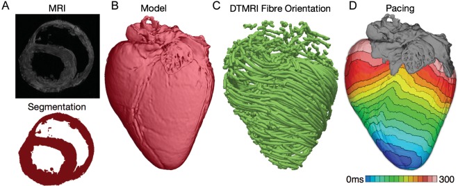
Figure 1.
Magnetic resonance imaging-based high-resolution model of electrical activation in the normal canine heart. (A) Segmentation of the myocardium. (B) Reconstructed canine heart geometry. (C) Fibre orientation in the canine heart as determined from the diffusion-tensor magnetic resonance imaging data. (D) Paced activation in the heart.

