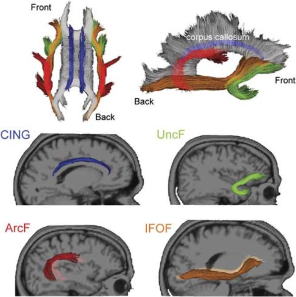
Fig. 1.

Fibers that project to the frontal lobe selected for the analysis. Top row shows the selected fibers from superior horizontal and mid-sagittal views. Middle and bottom rows present the individual fibers from sagittal view. CING appears in blue; UncF in green; ArcF in red and IFOF in orange.
