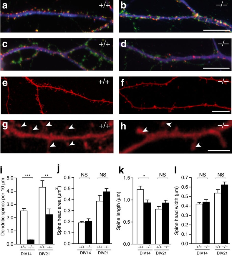
Figure 1. Nbea knockout neurons in culture fail to develop a normal number of dendritic spines.
(a,b) Immunocytochemistry of dendrites from wild-type (+/+) and Nbea-deficient (−/−) neurons co-labelled with antibodies against synapsin (green), PSD-95 (red) and MAP2 (blue) to visualize pre- and postsynaptic elements of excitatory synapses at DIV21. (c,d) Same experiment as in a,b but using antibodies against gephyrin (red) instead of PSD-95 to reveal inhibitory contacts. (e–h) Representative images of spine-bearing dendrites from wild-type and KO hippocampal neurons transfected with mRFP for 17 days, shown at lower magnification in a low-density culture56 (e,f), and at higher magnification in confocal images (g,h; arrowheads point to the spinous protrusions). (i–l) Histograms showing quantitative comparisons of spine density (i, numbers per dendrite length; data are from 7–9 independent cultures per genotype), and spine dimensions (j, spine head area; k, spine length; l, spine head width; data are from 4–6 independent cultures per genotype) between wild-type and Nbea-deficient neurons at two different time points (DIV14 and DIV21) in vitro. All data are means±s.e.m. *P<0.05, **P<0.001, ***P<0.0001, NS, not significant. Scale bars, 10 μm in a–f, 5 μm in g, h. DIV, days-in-vitro.

