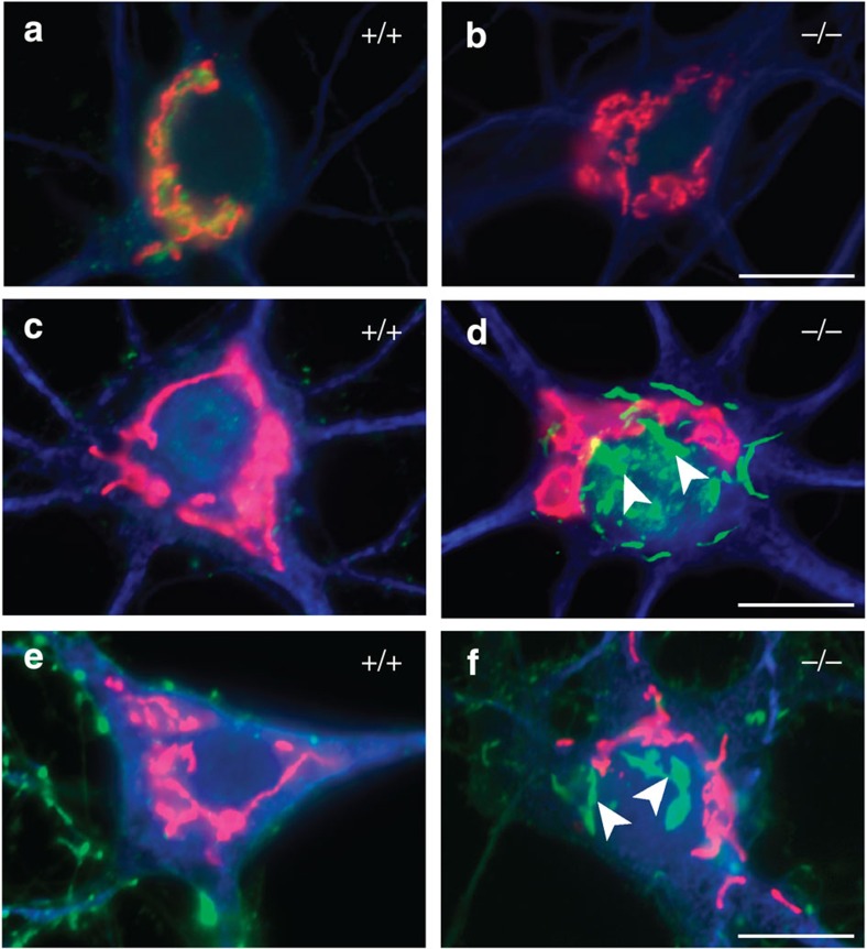
Figure 7. Synaptopodin and actin are retained near the trans-Golgi network in Nbea-mutant neurons.
(a) In wild-type (+/+) hippocampal neurons at DIV21, a large amount of Nbea (green) is localized near the trans-Golgi network revealed by the marker protein GM130 (red). Additional co-labelling against MAP2 (blue) identifies dendrites and somata. (b) Control experiment showing co-staining with the same antibodies in Nbea-deficient (−/−) hippocampal neurons. (c) Co-labelling of synaptopodin (green) with GM130 (red) and MAP2 (blue) fails to detect visible amounts of synaptopodin in somata of wild-type neurons. (d) In contrast, deletion of Nbea leads to ectopic accumulation of prominent clusters of synaptopodin (filled arrowheads) in the cell body near the trans-Golgi network. (e,f) Co-labelling of actin (green) with GM130 (red) and MAP2 (blue) reveals a comparable pattern to the synaptopodin/GM130 distribution (c, d), showing ectopic accumulation of actin clusters in the cell body of Nbea-deficient neurons (f, filled arrowheads). Scale bars, 10 μm in a–f.

