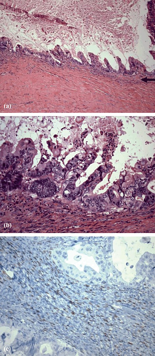
Figure 3.

Histopathology showing (a) the cyst lining with papillary projections into the cyst lumen and ovarian-type stroma (arrow) and (b) high-grade dysplasia/carcinoma in situ overlying spindle cell/ovarian stroma. [Haematoxylin and eosin stain; original magnification (a) ×10, (b) ×40.] (c) Immunostain for oestrogen receptor highlighting ovarian-type stroma. (Original magnification ×40)
