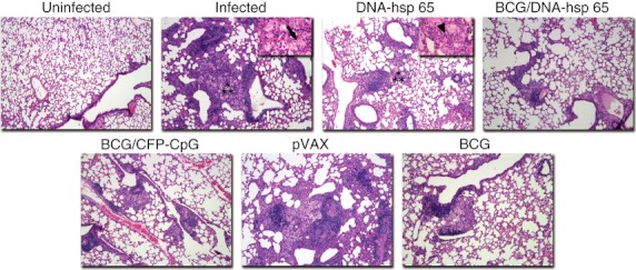
Figure 3.

Histological analysis of lungs. Mice were vaccinated with DNA-heat-shock protein (hsp) 65, BCG/DNA-hsp 65 or BCG/CFP-CpG and challenged with Mycobacteirum tuberculosis as described in Fig. 2. Pathology was evaluated on the upper right lobe of the lung on day 30 post-challenge (original magnification, 100 ×). Asterisks show the areas represented in 400 × magnification (upper right quadrant). Arrow shows a foamy macrophage. Head arrow shows neutrophils.
