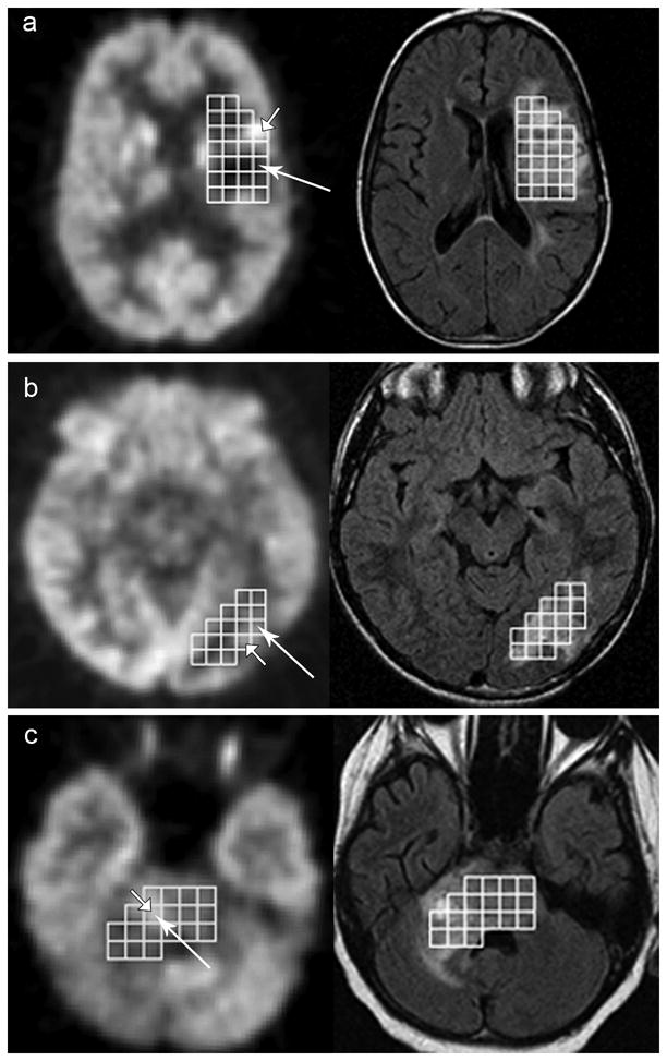
Figure 1. Voxel-wise comparison of FDG-PEG and MRSI.

FDG-PET (left) and FLAIR (right) images with the MRSI ROI voxels for 3 patients on study. Brightest voxel on PET indicated by short arrow. Voxel with Maximum Cho:NAA indicated by long arrow. A) No agreement between MRSI and PET voxel selection. B and C) Agreement between MRSI and FDG-PET. B) Selected voxels are adjacent to one another and C) the same voxel was chosen by both FDG-PET and MRSI.
