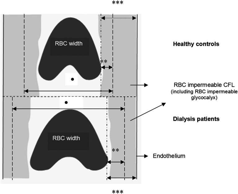
Figure 4.
Schematic illustration of endothelial glycocalyx imaging method. In dialysis patients, perturbation of glycocalyx allows the erythrocytes to approach the vessel wall, leading to increased DPerf and PBR compared with healthy controls. RBC width, median RBCW. •DPerf is the perfused diameter (RBC perfused lumen). **PBR is the perfused boundary region (RBC-permeable part of the cell-free layer including cell-permeable glycocalyx). ***Cell-free layer.

