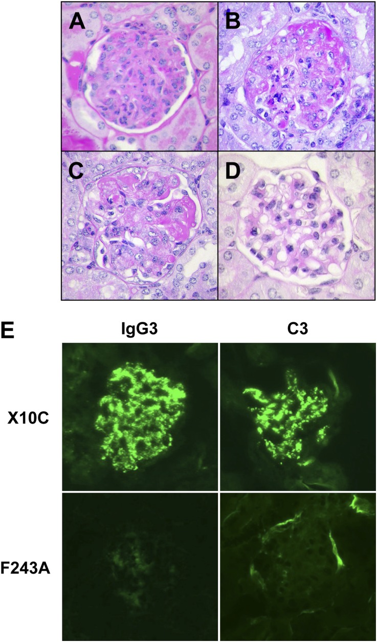
Figure 3.
Induction of glomerular lesions in BALB/c mice implanted with X10C or 6-19.ST6-5 IgG3 RF-secreting cells, but not with 6-19 F243A-secreting cells. (A and B) Representative glomerular appearance of early and more advanced lesions in BALB/c mice implanted with X10C hybridoma. Note infiltrations by PMN, proliferation of mesangial cells, expansion of mesangial matrix, as well as the marked deposition of PAS-positive materials in glomeruli. (C) Representative glomerular lesions of BALB/c mice implanted with 6-19.ST6-5 hybridoma. (D) Representative appearance of glomeruli in BALB/c mice implanted with 6-19 F243A transfectoma. Note essentially normal histologic appearance of glomeruli. (E) Immunohistochemical analysis of glomerular lesions of BALB/c mice implanted with X10C or 6-19 F243A-secreting cells. Note fine granular deposits of IgG3 and C3 in the mesangium and glomerular capillary walls in mice implanted with X10C cells, and the lack of significant immune deposits in mice implanted with 6-19 F243A cells. PAS staining in A–D. Original magnification, ×400 in A–D; ×200 in E.

