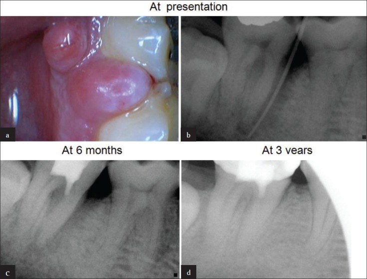
Figure 1.

(a) A 53-year-old man presented with discrete soft tissue swellings at gingival margin in relation to # 47. (b) Sinus tract traced with guttapercha point documenting a pulpoperiodontal lesion. Note the bone loss on mesial aspect of mesial root. (c) Follow up X-ray at 6 months showing healing of periodontal defect. (d) At 3-years, complete healing and new bone regeneration in inter dental area between #46 and #47 is evident
