
Figure 2.
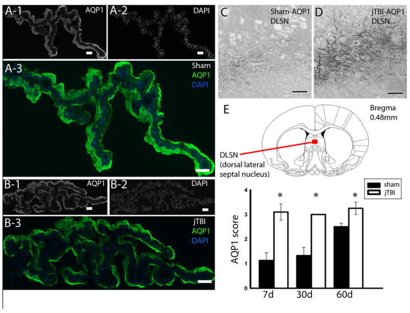
(A) AQP1 staining in the choroid plexus of sham and (B) jTBI shows no difference in intensity or pattern of staining. (C) AQP1 staining in the dorso-lateral septal nucleus of sham compared to that of (D) jTBI shows a (E) significant increase in staining in the jTBI animals at 7d, 28d, and 60d.
(AQP1,aquaporin1; jTBI, juvenile traumatic brain injury; DLSN, dorsal lateral sepal nucleus; d,days post injury; scale bar = 100 μm, *p<0.01).