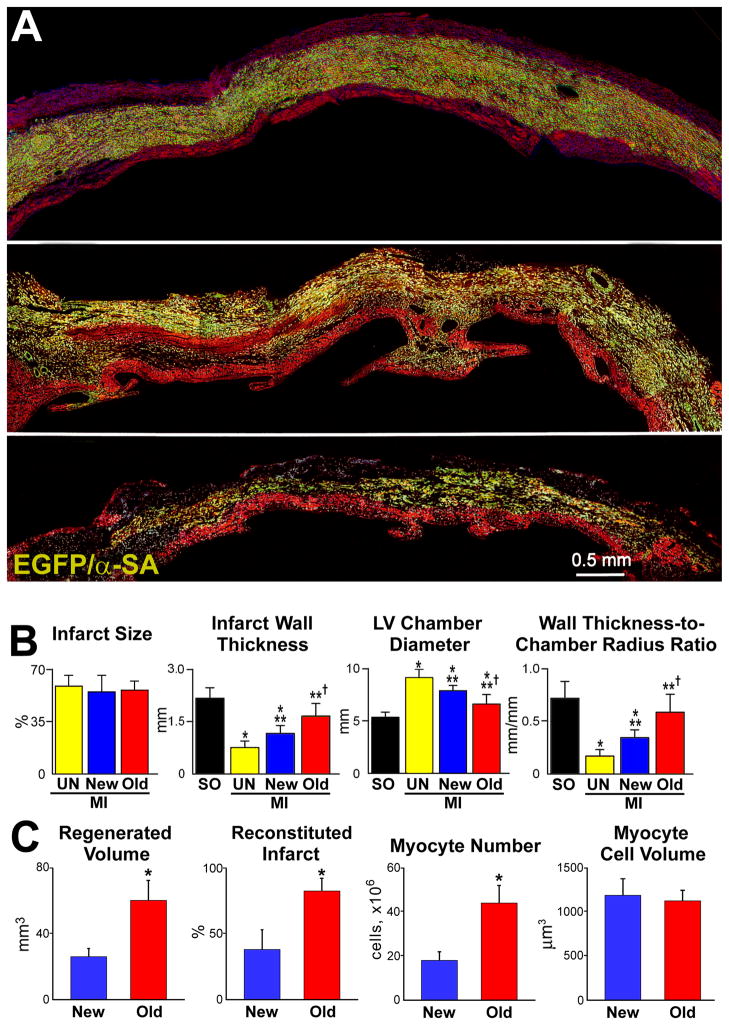
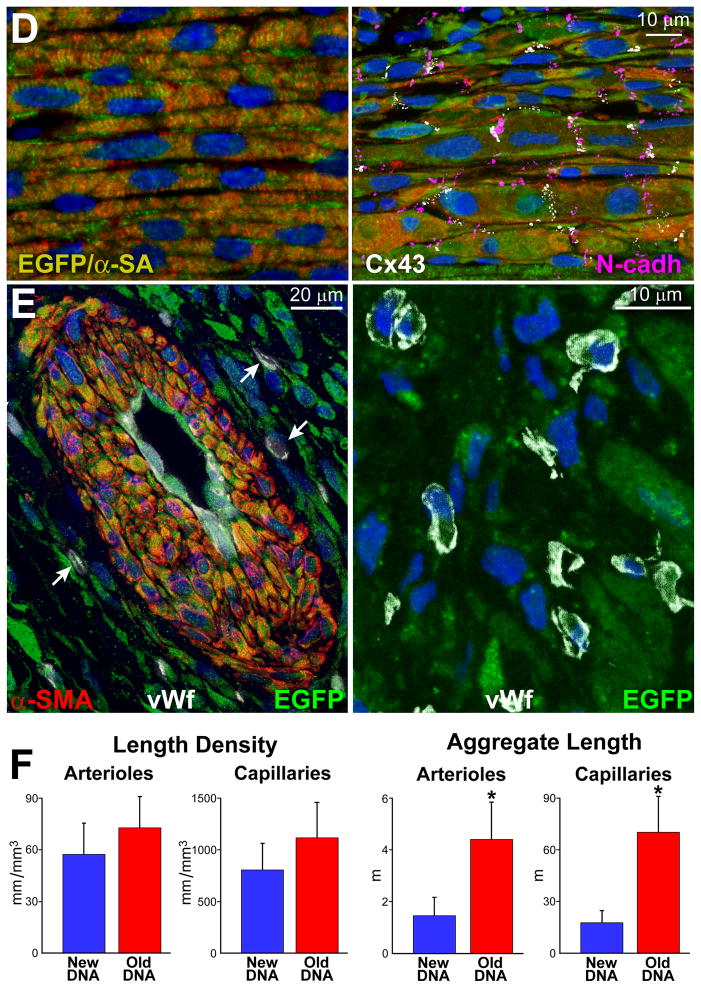
Figure 6. Myocardial regeneration and hCSC classes.
A, hCSCs carrying the old DNA reconstitute most of the infarct (upper and central: combination of EGFP and α-SA, yellowish. Labeling by EGFP, α-SA, and merge are shown in Online Figure XA through XC. hCSCs carrying the new DNA replace only in part the infarcted myocardium (lower). B, Infarct size is similar in all cases. hCSCs obtained by FACS-FRET or UV light produced similar results, which were combined. hCSCs with old DNA have a more positive effect on the thickness of the infarcted wall, chamber diameter, and wall thickness-to-chamber radius ratio than hCSCs carrying the new DNA. SO, sham-operated; MI, infarct; UN, untreated; New, treated with hCSCs with new DNA; Old, treated with hCSCs with old DNA. Values are mean±SD. *,**,†P<0.05 vs. SO, UN, and New, respectively. C, The amount of regenerated human myocardium is 2.4-fold larger with hCSCs with old DNA, reconstituting most of the infarcted wall; this was mediated by a 2.5-fold increase in the number of newly-formed myocytes of comparable size. *P<0.05 vs. New. D, Human myocytes are EGFP-positive and show sarcomere striation: EGFP and α-SA (yellowish). Human myocytes express the junctional proteins connexin 43 (Cx43, white) and N-cadherin (N-cadh, magenta). E, Human arteriole is EGFP-positive and is composed of several layers of SMCs (combination of EGFP and α-SMA, yellowish) and a thin layer of ECs (combination of EGFP and vWf, light green); EGFP, α-SMA, vWf, and merge are shown in Online Figure XD. Similarly, human capillaries are EGFP-positive and are composed of a thin layer of ECs (combination of EGFP and vWf, light green). F, Length density and aggregate length of arterioles and capillaries in the regenerated myocardium with hCSCs carrying the old and new DNA. *P<0.05 vs. New.


