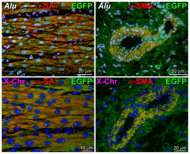
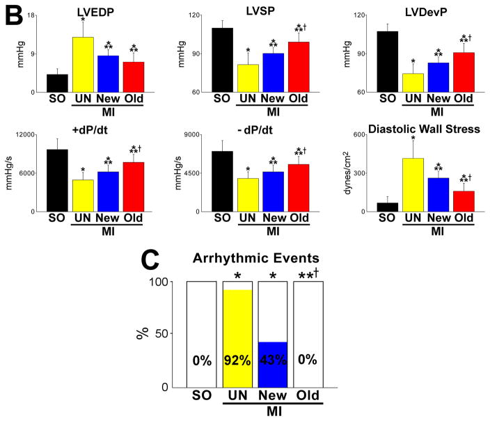
Figure 7. Human structures and ventricular function.
A, Human myocytes and vessels show human DNA sequences (Alu probe, white dots in nuclei) and human X-chromosome (X-Chr, single magenta dots in nuclei). B, hCSCs with old DNA had a more positive effect on left ventricular (LV) end-diastolic pressure (LVEDP), LV systolic pressure (LVSP), LV developed pressure (LVDP), positive and negative dP/dt, and calculated diastolic wall stress than hCSCs carrying the new DNA. C, Number of arrhythmic events in SO, and untreated and treated infarcts. For abbreviation and statistics see Figure 6B.


