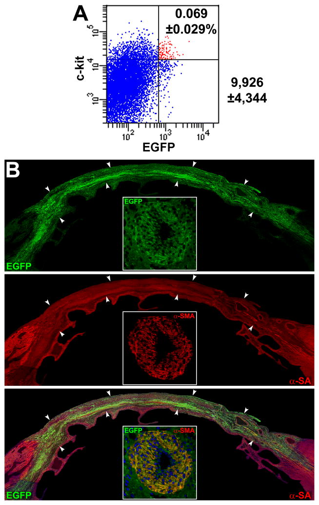
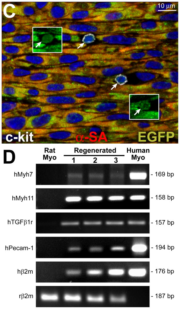
Figure 8. Serial transplantation of clonal hCSCs with old DNA.
A, c-kit-positive EGFP-positive hCSCs (red dots) isolated after regeneration of the infarcted heart are shown by scatter plot. B, Following serial transplantation, a large area of the infarcted myocardium is replaced by EGFP-positive (upper, green), α-SA-positive (central: red, arrowheads) cardiomyocytes (lower: merge, yellowish, arrowheads). A coronary vessel (inset) is positive for EGFP (upper, green), and α-SMA (central, red); merge (lower, yellowish). C, In a serially transplanted infarcted heart, EGFP-positive (insets) c-kit-positive (white) hCSCs are present (arrows). D, Expression of human genes by q-RT-PCR in cell treated infarcted hearts: cardiomyocyte (β-myosin heavy chain, hMyh7), SMC (SM-myosin-heavy-chain 11, hMyh 11; TGF-β1 receptor, hTGFβ1r), and EC (hPecam-1) genes are shown together with the housekeeping gene human β2-microglobulin (hβ2m) and rat β2-microglobulin (rβ2m). Rat and human myocardium (Myo) were used as negative and positive control, respectively. The PCR products had the expected molecular weight. For tracings and sequences of PCR products, see Online Figure XI.


