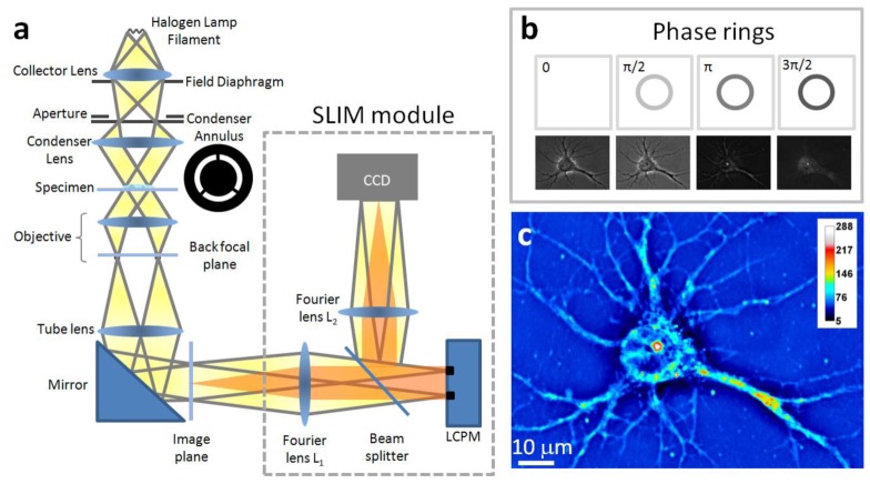
Fig. 1.
SLIM principle. (a) Schematic setup for SLIM. The SLIM module is attached to a commercial phase contrast microscope (Axio Observer Z1, Zeiss, in this case). The lamp filament is projected onto the condenser annulus. The annulus is located at the focal plane of the condenser, which collimates the light towards the sample. For conventional phase contrast microscopy, the phase objective contains a phase ring, which delays the unscattered light by a quarter wavelength and also attenuates it by a factor of 5. The image is delivered via the tube lens to the image plane, where the SLIM module processes it further. The Fourier lens L1 relays the back focal plane of the objective onto the surface of the liquid crystal phase modulator (LCPM, Boulder Nonlinear). By displaying different masks on the LCPM, the phase delay between the scattered and unscattered components is modulated accurately. Fourier lens L2 reconstructs the final image at the CCD plane, which is conjugated with the image plane. (b) The phase rings and their corresponding images recorded by the CCD. (c) SLIM quantitative phase image of a hippocampal neuron.

