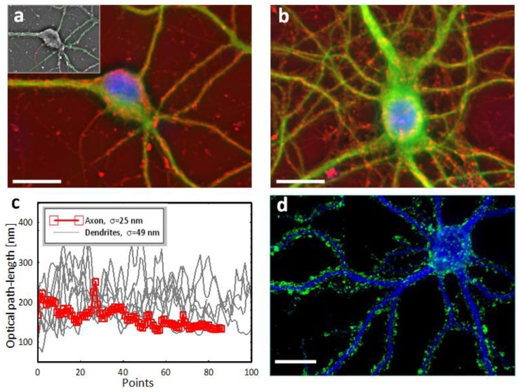
Fig. 4.
SLIM-fluorescence multimodal imaging. (a)-(b) Combined multimodal images of cultured neurons (19 DIV) acquired through SLIM (red) and fluorescence microscopy of anti-MAP2 stained soma and dendrites (green) and DAPI-stained nuclei (blue). (c) Optical path-length fluctuations along the dendrites (green) and axon (red) retrieved from the inset of (a). (d) Synaptic boutons of a mature hippocampal neuron (33 DIV) immunochemically labeled for synapsin (green) and MAP2 (blue). Scale bars: 20 μm.

