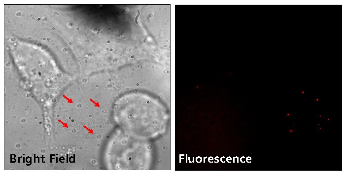
Fig. 5.

Bright field (left) and fluorescence (right) images of the HEK cells cultured on the fiduciary-marked coverslip with quantum dots (605 nm emission; Invitrogen) labeling. In the bright-field image, there are cultured cells as well as regular patterned dots (represented by arrows) which are the fiduciary markers. The fluorescence image has no autofluorescence, showing clear emission spots of the qdots.
